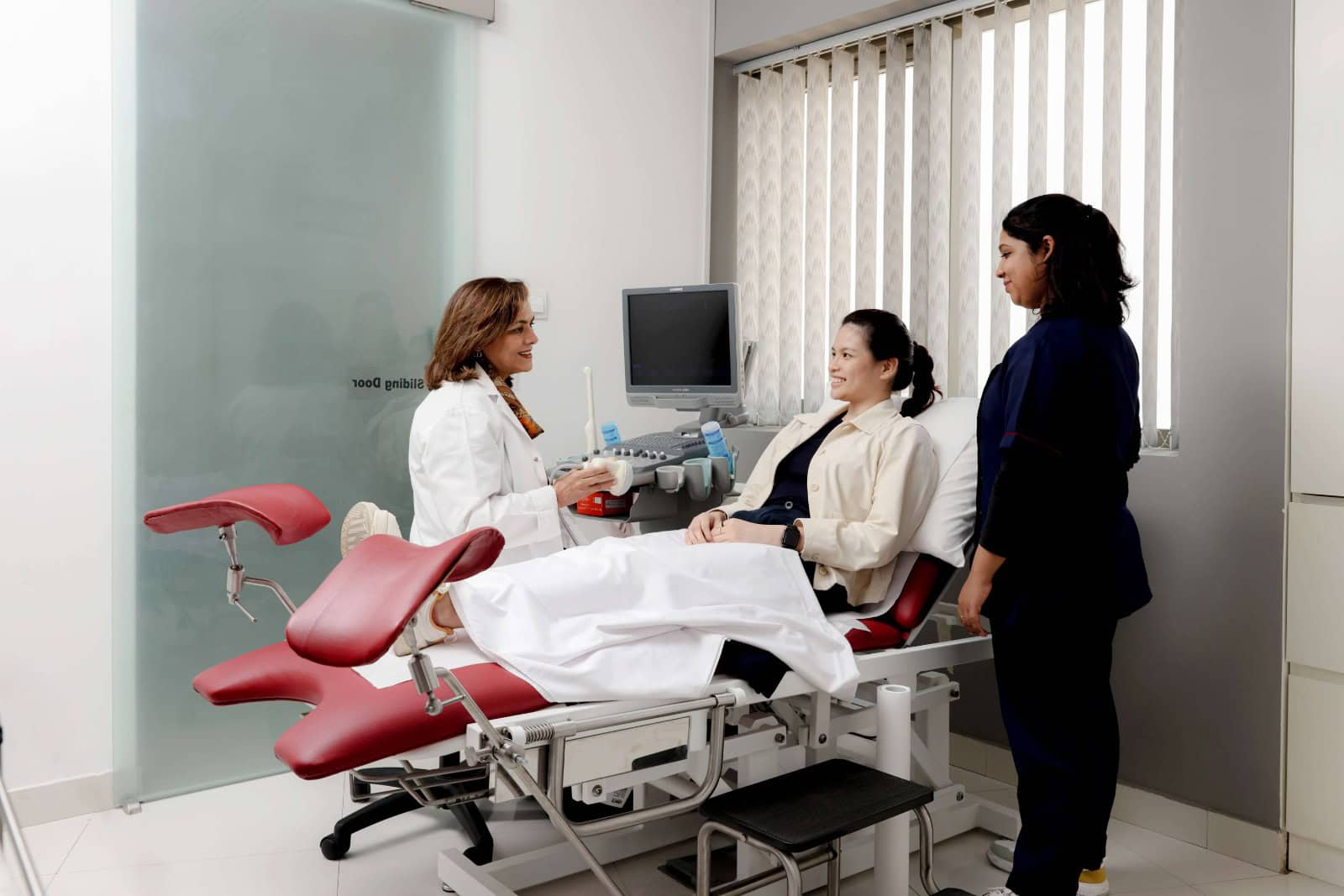
Internal signals like sudden weight shifts or persistent breakouts are the body’s way of requesting an investigation. When cycles become unpredictable, the biological rhythm needs recalibrating through diagnostics.
Modern imaging offers a window into these hormonal complexities, providing the data necessary to restore balance. For imaging and a detailed assessment, you need to visit a dedicated female gynecologist in Dubai.
The string of pearls
When a doctor moves the ultrasound wand, they search for a very specific pattern on the ovaries. In many cases, they see several small fluid filled sacs resting near the surface. These are actually follicles that did not fully develop or release an egg. On the screen, they look like tiny black circles. Because they often sit in a row, doctors frequently call this the string of pearls appearance.
Ovary size and volume
A healthy ovary is usually about the size of an almond. During the scan, the doctor measures the length, width, and depth of each one. Ovaries affected by this condition often appear larger than normal. This happens because the extra follicles and dense tissue take up extra space. Seeing enlarged ovaries helps the medical team confirm that the hormone levels are impacting the physical shape of the reproductive organs.
The thick outer layer
The outer edge of the ovary, known as the cortex, can tell a story of its own. In some patients, this outer shell looks much thicker or denser than expected. This thickness can sometimes make it harder for eggs to be released during a normal cycle. The ultrasound allows the doctor to see this density clearly.
Blood flow patterns
Modern ultrasound machines can also check how blood moves through the pelvic area. Doctors look at the vessels surrounding the ovaries to see if the flow is higher than usual. Increased blood movement in specific spots can be a sign of high activity or hormonal shifts. This data adds another layer of proof to the diagnosis and helps rule out other health concerns.
Checking the uterine lining
The scan is not simply about the ovaries. The doctor also looks at the lining of the uterus. Since cycles are often irregular with this condition, the lining might become quite thick. Seeing the thickness of this tissue helps the doctor decide on the best way to manage the cycle. It ensures that the uterus is healthy and helps prevent future complications related to infrequent periods.

